ArnoldChiari malformation CT wikidoc
Not everyone with a Chiari malformation requires surgery, but when a patient's individual circumstances warrant it, a neurosurgeon may recommend "decompression" surgery (known as a "decompressive suboccipital craniectomy and cervical laminectomy").
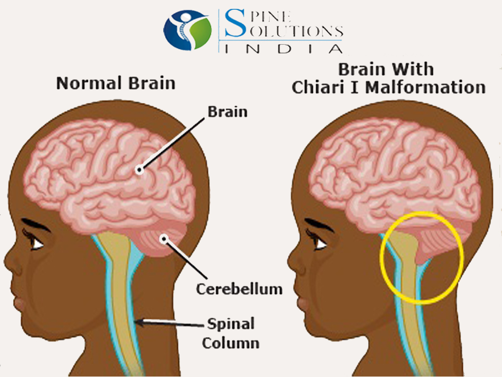
Spine Solutions India by Dr Sudeep Jain Chiari Malformation A structural defect in the Cerebellum
Chiari Malformation is a serious neurological disorder where the bottom part of the brain, the cerebellum, descends out of the skull and crowds the spinal cord, putting pressure on both the brain and spine and causing many symptoms.. Latest Chiari Research. Tonsil Resection May Be More Effective Than Duraplasty For Chiari Related.
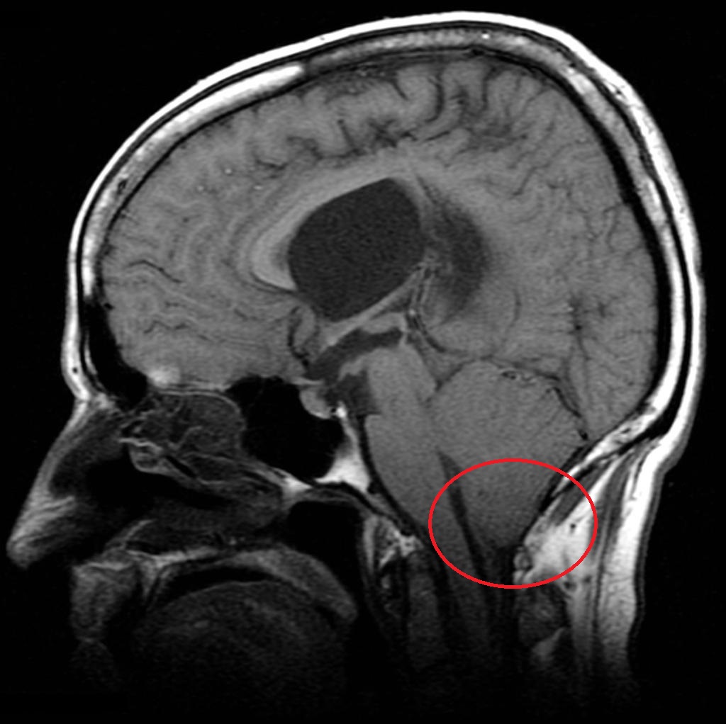
Arnold Chiari Type I Malformation C.N.S. Neurosurgery
Check out our chiari malformation zipperhead selection for the very best in unique or custom, handmade pieces from our laptop decals shops.

Zipper head Chiari malformation, Chiari, Awareness
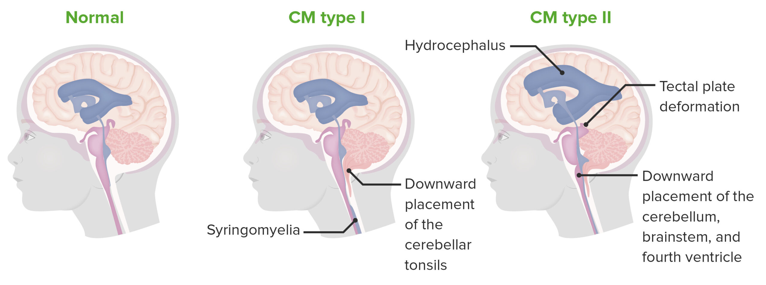
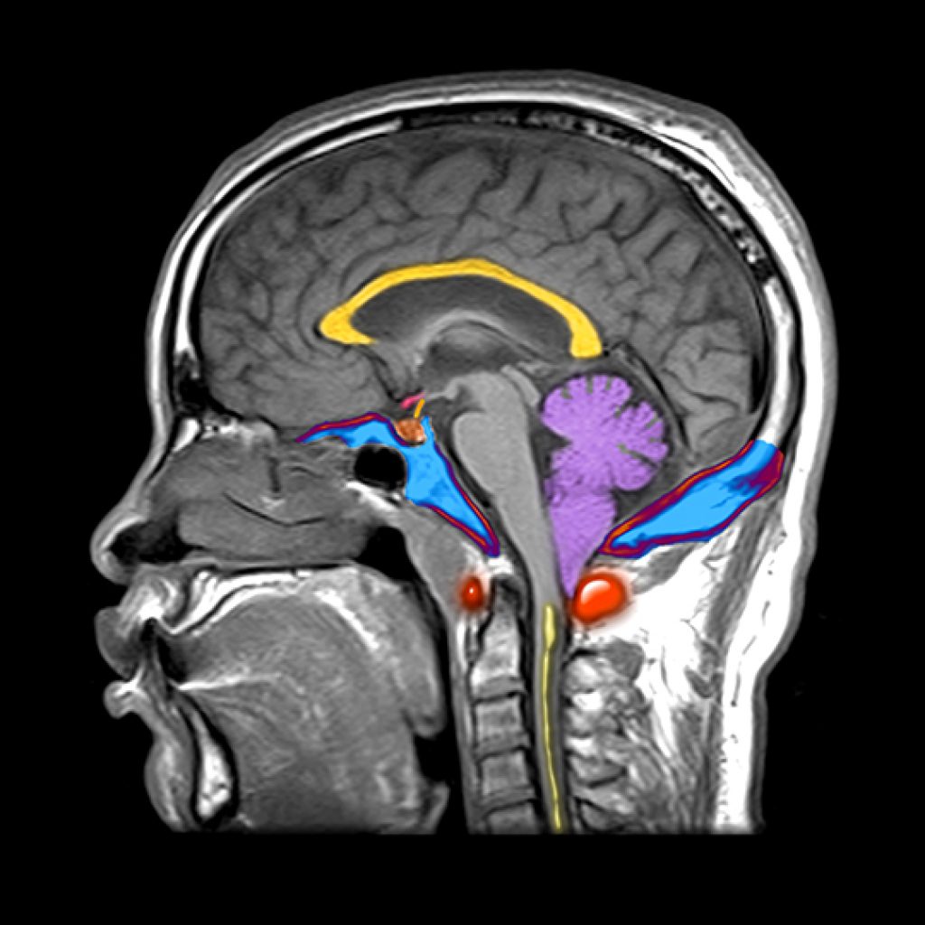
Chiari malformations are structural defects in the base of the skull and cerebellum, the part of the brain that controls balance and movement. Normally, the cerebellum and parts of the brainstem sit above a natural opening at the base of the skull called the foramen magnum, which allows the spinal cord to pass through the skull..
Chiari Malformations Neurosurgery Geeky Medics
Chiari malformation is considered a congenital condition, although acquired forms of the condition have been diagnosed. In the 1890s, a German pathologist, Professor Hans Chiari, first described abnormalities of the brain at the junction of the skull with the spine. He categorized these in order of severity; types I, II, III and IV.

Chiari Malformations Neurosurgery Geeky Medics
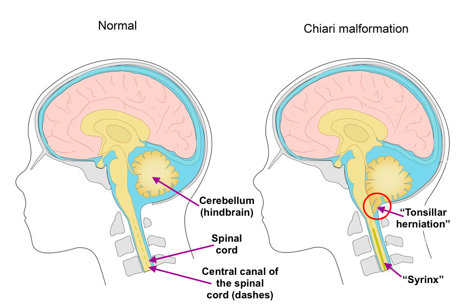
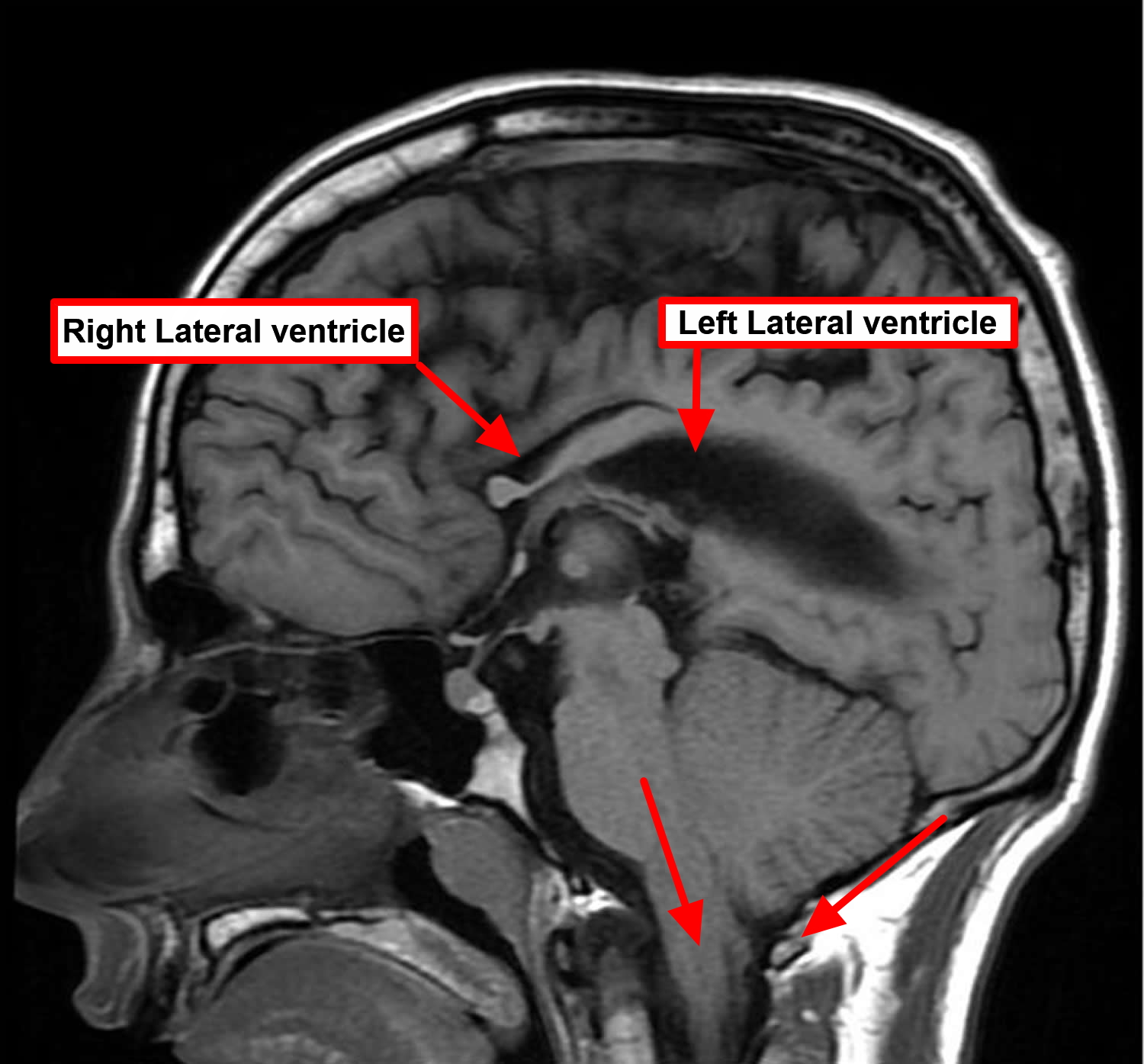
Chiari disease (or malformation) is in general a congenital condition characterized by an anatomic defect of the base of the skull, in which the cerebellum and brain stem herniate through the foramen magnum into the cervical spinal canal. The onset of Chiari syndrome symptoms usually occurs in the second or third decade (age 25 to 45 years). Symptoms may vary between periods of exacerbation.

Pin on Chiari Malformation
Chiari malformations (CM) are caused by problems in the structure of the brain and skull. In Chiari malformations, the lower part of the brain presses on and through an opening in the base of the skull and cerebellum into the spinal canal. The cerebellum is the part of the brain that controls balance.
Chiari Malformations Concise Medical Knowledge
Chiari malformations are a heterogeneous group of hindbrain anomalies. Six different malformations are described. Most common are Chiari 1 malformation (CM1) and Chiari 2 malformation (CM2, also termed "Arnold-Chiari malformation") and are the focus of this review. These are rare conditions, but symptoms may impair quality of life in both.

Pin de Lauren VanHoose en chiari Tatuaje para cubrir cicatriz, Tatuajes de portada, Tatuaje en
Chiari Zipperhead Decal | Chiari Malformation | Arnold Chiari | Spoonie (3.7k) $3.25 FREE shipping Brain surgery survivor - Chiari malformation shirt - ACM awareness - zipperhead shirt - brain sweatshirt - Chiari shirt - purple awareness (2k) $25.00 20 oz Chiari Warrior Tumbler (1k) $25.00
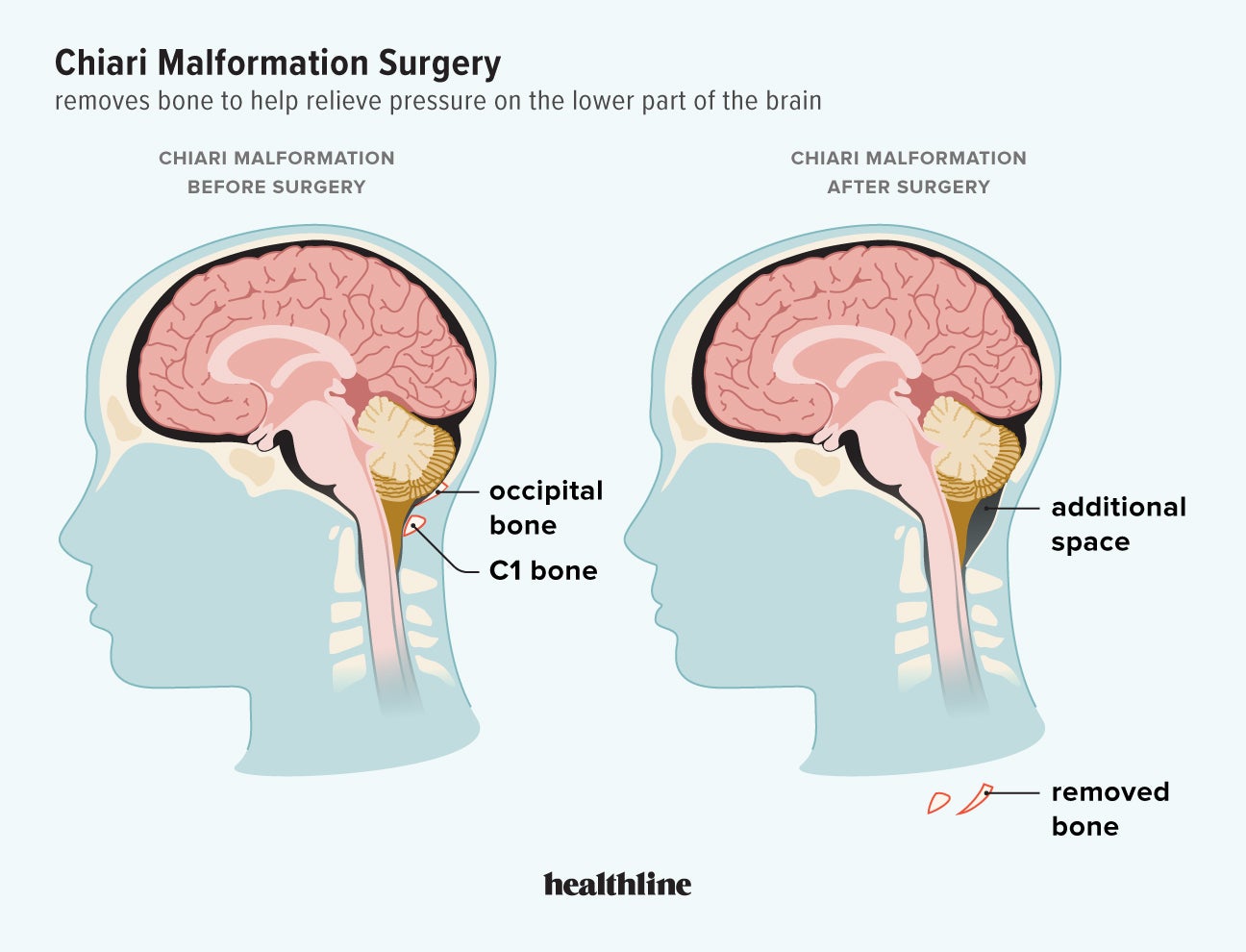
Chiari Malformation Surgery Candidates, Procedure, Recovery, and More
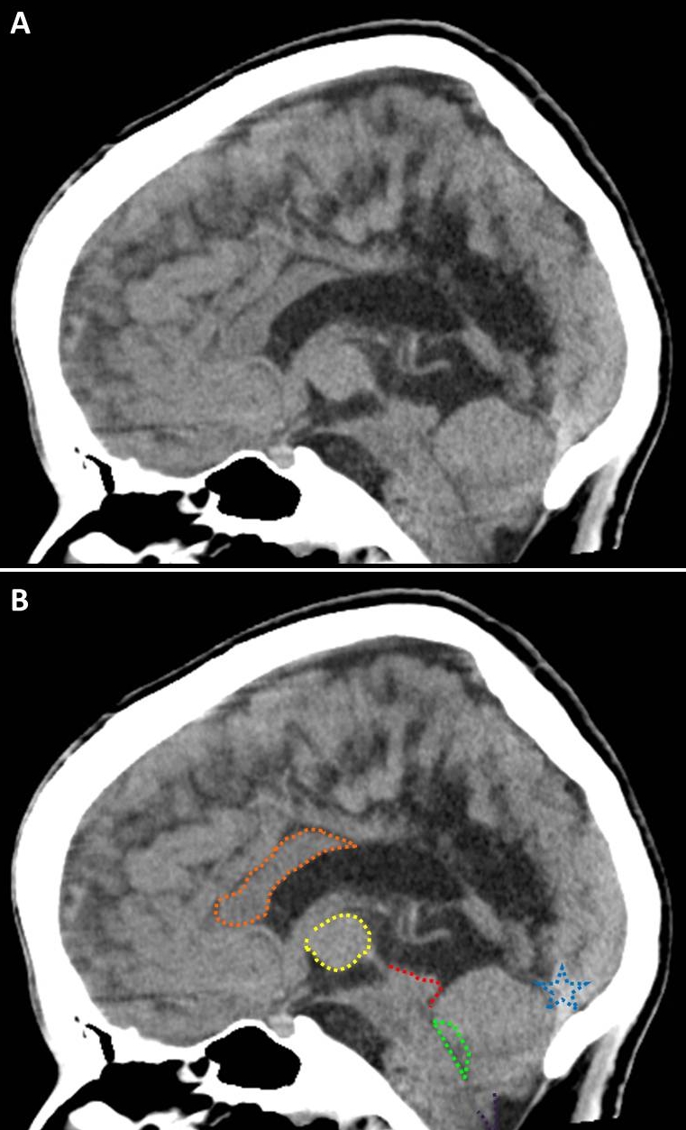
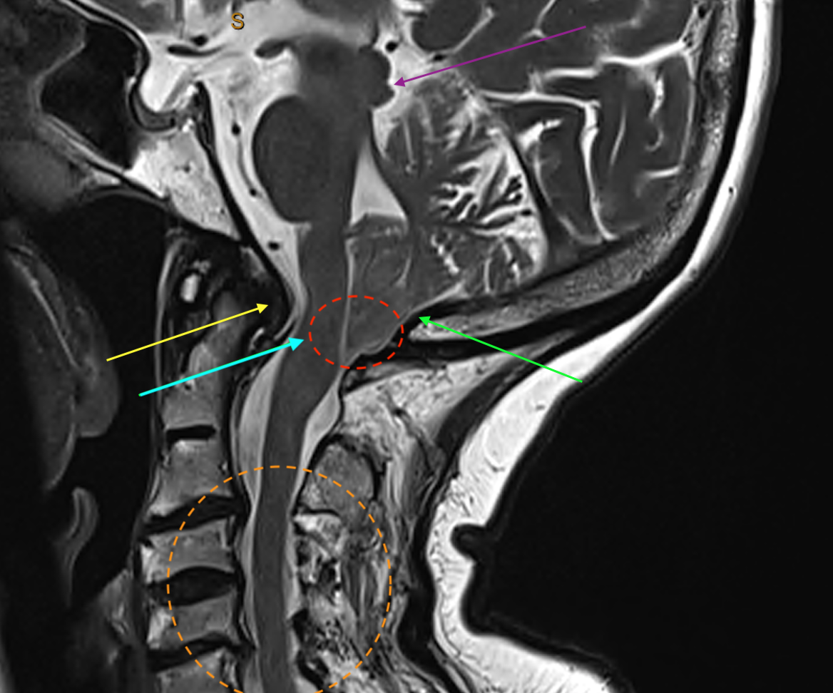
The Chiari I malformation is characterized by an inferior position of the cerebellar tonsils relative to the foramen magnum. This is believed to be due to a mismatch between the size and content of the posterior fossa. Four groups of Chiari I patients can be distinguished, according to different pathogeneses 9:
Chiari Malformation Causes, Symptoms, Prognosis, Diagnosis, Treatment
Chiari malformation (kee-AH-ree mal-for-MAY-shun) is a condition in which brain tissue extends into the spinal canal. It occurs when part of the skull is misshapen or smaller than is typical. The skull presses on the brain and forces it downward. Chiari malformation is not common, but increased use of imaging tests has led to more diagnoses.

I'm a zipper head. Medical education, Chiari malformation, Medical
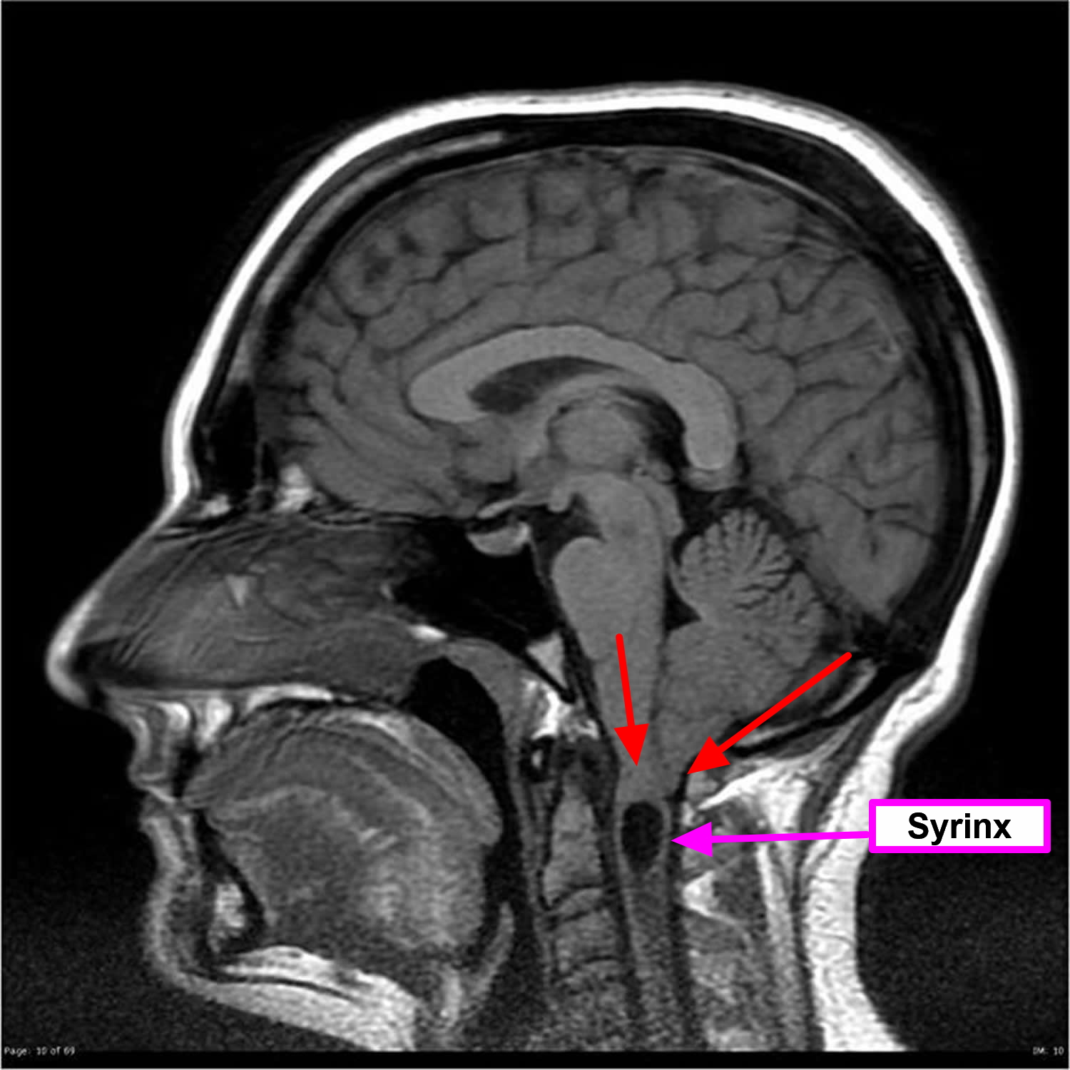
Synopsis. Chiari Malformation Type I (CMI) is a congenital malformation diagnosed by MRI findings of at least 5 mm of cerebellar ectopy below the foramen magnum. CM1 is frequently associated with syringomyelia. Herein, we discuss the history of CMI and syringomyelia, including early pathologic and surgical studies.
Arnold Chiari Type I Malformation C.N.S. Neurosurgery
Chiari II malformation (CM-II), commonly known as Arnold-Chiari malformation, is a relatively common congenital malformation characterized by beaked midbrain, downward displacement of the tonsils, and cerebellar vermis, and spinal myelomeningocele.[1] This malformation is frequently misunderstood as a more severe version of Chiari I malformation (CM-I). However, these are two distinct diseases.
Chiari Malformation Neurosurgery and Endovascular Associates
Chiari malformation is when a part of the brain extends through an opening where the skull meets the spinal canal. It can happen if the skull is too small or misshapen. Depending on the severity.

Pin on Chiari malformation
A Chiari malformation can also cause pressure on the brain and produce hydrocephalus (pressure due to excessive cerebrospinal fluid accumulation in the brain) and the spinal cord, potentially causing a wide variety of symptoms. In fact, no two cases of Chiari malformation are exactly alike and the associated symptoms are highly variable.
Chiari Malformation Causes, Symptoms, Prognosis, Diagnosis, Treatment
Pathogenesis. There is increasing evidence that Chiari malformations are due to underdevelopment of the posterior cranial fossa, resulting in overcrowding compared with the normally developed hindbrain. 1,2 The posterior cranial fossa is the part of the cranial cavity, which contains the cerebellum and lower brainstem (ie, the pons and the medulla). A smaller cranial fossa leaves a typically.